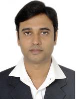
Dr. Vishal Srivastava
Dr. Vishal Srivastava received his Ph.D. degree at Indian Institute of Technology Delhi Instrumentation Design and Development Center. He has more than 10 years of teaching, research and Industrial experience. He has authored two book chapters, and published more than 28 papers in international refereed journals and conferences.
| Phone/Mobile Number | 8198051071 |
| Email ID | vsrivastava@thapar.edu |
“Development of full-field polarization sensitive optical coherence tomography for soft biological tissue” Approved by DST-SERB
IEEE, SPIE
Azeem Ahmad, Vishal Srivastava, Vishesh Dubey, and D.S. Mehta, “Ultra-shot longitudinal spatial coherence length of laser light with combined effect of spatial, angular and temporal diversity” Applied Physics letter,Vol.106,093701 (2015).
Anuj Kumar, Vishal Srivastava, Manoj Kumar Singh, GP Hancke, “Current Status of the IEEE 1451 Standard Based Sensor Applications”,IEEE Sensors, Issue 99, (2014).
Vishal Srivastava, Sreyankar Nandy and Dalip Singh Mehta,“High-resolution full-field opticalcoherence tomography using a spatially incoherent monochromatic light source”, AppliedPhysics letter, Vol.103, 103702 (2013).(Also appeared inOCT news,http://www.octnews.org/articles/4858678/high-resolution-fullfield-spatial-coherence-gated/)
Vishal Srivastava, Sreyankar Nandy and Dalip Singh Mehta,“High-resolution cornealtopography and tomography of fish eye using wide field white light interference microscopy”,Applied Physics letter, Vol.102, 153701 (2013).(Also appeared in OCT news, http://www.octnews.org/articles/4540585/high-resolution-corneal-topography-and-tomography-/)
Dalip Singh Mehta and Vishal Srivastava, “Quantitative phase imaging of human red blood cells using phase-shifting white light interference microscopy with colour fringe analysis”, Applied Physics letter,Vol.101, 203701 (2012). (Also appeared in OCT news, http://www.octnews.org/articles/4550444/quantitative-phase-imaging-of-human-red-blood-cell/)
Full-field optical coherence tomography and microscopy using spatially incoherent monochromatic light, Dalip Singh Mehta, Vishal Srivastava, Sreyankar Nandy, Azeem Ahmad and Vishesh DubeyHandbook of Optical Coherence Microscopy Technology and Applications, Volume 1, CRC Press Taylor and Francis Group
White Light Phase-Shifting Interference Microscopy for Quantitative Phase Imaging of Red Blood Cells, Dalip Singh Mehta &Vishal Srivastava, pp 581-584, Springer.
Received Best Poster award in WRAP 2013 Workshop on Recent Advances in Photonics, IIT Delhi, India December 17-18 (2013).
Received second best Project award during the I2 Tech – Open House held on 20th April 2013 in Indian Institute of Technology Delhi.
Awarded travel grant from Department of science and technology (DST), India to participate the “SPIE Photonics West conference on BIOS,” San Francisco, California, Feb02-07, (2013).
Received Best Poster award in XXXVI Optical Society of India Symposium Frontiers in Optics and Photonics (FOP11), IIT Delhi, India December 2-5 (2011).
Awarded MHRD scholarship for 2009- 2013; for the Ph. D. duration
Awarded MHRD scholarship for 2005- 2007; for the M. Tech. duration.
GATE qualified in Electronics and Communication Engineering in 2004 and 2005.
His area of research includes signal processing, Image processing, development of biomedical instruments, wide field imaging and health monitoring. He developed three biomedical instruments for quantitative phase imaging of biological cells. His research contributions are in Biophotonics and Biomedical Optics and development and application of various optical methods for non-invasive and non-destructive imaging of biological cells He is interested in the development of biomedical devices and image processing applications.